Unlocking the Power of Cortical Plasticity
Introduction
Cortical plasticity and neuroplasticity are terms that are often used to mean the same thing. They both describe how the brain can create new pathways for sending nerve signals as a reaction to changes inside the body or to its environment (1). These new brain pathways lead to changes that help the brain adapt and even promote the recovery of lost skills in stroke patients.
The brain has three main parts: cerebrum, cerebellum, and the brainstem. The cerebrum’s outer layer, called the cortex, is made of gray matter and makes up about half of the brain’s weight (2). Cognitive functioning, or everyday thinking and mental activities, are mostly managed by the cortex and the deeper parts of the cerebrum (3).
The following article will describe how stroke damage to the different parts of the cerebrum can result in problems with motor skills and cognitive functioning, as well as how cortical plasticity boosts stroke recovery. The article will also explore how occupational and physical therapy (OT and/or PT) incorporates cortical plasticity into their therapeutic approaches, and the strategies that stroke survivors can utilize cortical plasticity to boost their stroke recovery.
Brain Hemispheres: Regions, Functions, and Effects of Stroke
The brain’s left and right hemispheres each control different functions, and damage to specific lobes in a hemisphere can affect abilities like speech, memory, and vision.
The cerebrum has a left and a right hemisphere. The left hemisphere is typically dominant in a right-handed person, and vice versa. A person’s dominant hemisphere typically controls speech and language comprehension (4).
Each hemisphere is further composed of the following parts, or lobes (5):
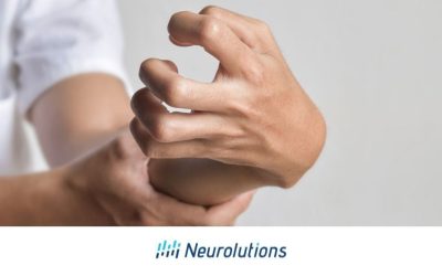
- Frontal lobe: This is the largest lobe and is found at the front of both hemispheres of the brain. If it gets damaged, short-term memory, personality, and speech can be affected. The Broca area, located in the frontal lobe, controls speech production. If someone suffers damage to their Broca area from a stroke, they often have trouble speaking.
- Parietal lobe: This lobe is located behind the frontal lobe and above the temporal lobe. It plays a key role in interpreting sensory information like touch, vibration, pain, and temperature. It also helps with understanding visual and auditory information, learning, planning movements, spatial awareness, and other skills needed for everyday activities.
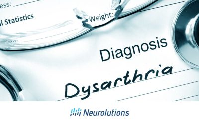
- Temporal lobe: This lobe is located behind the frontal lobe and underneath the parietal lobe. It controls the interpretation of auditory input, and it also contains the Wernicke’s area. The Wernicke’s area is responsible for language comprehension. If this area suffers damage from a stroke, it can make it difficult for a survivor to read, write, and understand spoken language. Additionally, damage to the temporal lobe can impair a person’s ability to recognize objects and remember events.
- Occipital lobe: This is the smallest lobe and is located at the back of the cerebrum. The occipital lobe controls vision, and if it is damaged, a person may go blind even if their eyes are healthy. Damage to the occipital lobe can also result in visual processing issues, such as difficulty recognizing objects, faces, or interpreting visual information.
Understanding Cortical Plasticity and Its Role in Stroke Recovery
Cortical plasticity allows undamaged brain areas to take over lost functions after a stroke, aiding in recovery of motor and cognitive skills.
Neuroplasticity, also known as cortical plasticity, helps other parts of the brain take over lost functions after a stroke. This means that undamaged areas can learn to handle the tasks that were previously managed by the damaged parts. Recovery of cognitive skills, such as short-term memory, can also be improved if rehabilitation efforts are focused on refining that specific lost skill.
Recovery is more likely to occur in people with mild or moderate strokes because it’s more difficult for the brain to develop new pathways after a widespread or severe stroke. Additionally, using cortical plasticity to regain lost skills requires attention and focus, which might be significantly impaired after a severe stroke (6).
The concept of plasticity has been known since the late 19th century. Today, most healthcare providers understand that “re-wiring” the brain is a key part of recovering motor skills, alongside OT and/or PT (7, 8). Medical researchers are able to track the development and progress of cortical plasticity over time in stroke survivors using MRI scans (9). EEG can be a valuable tool in detecting and studying cortical plasticity by observing how brainwave patterns evolve in response to various stimuli or rehabilitation efforts.
This re-wiring is further enhanced by participating in OT and/or PT focused on motor skill recovery. As motor skill recovery progresses, cognitive skills might also improve. This means that a stroke survivor that is focused on relearning to self-feed and self-dress can also experience an improvement in attention, concentration, spatial awareness.
Neurorehabilitation Strategies for Enhancing Brain Plasticity
Therapies focused on attention and motor skills, like OT and PT, help stroke survivors improve cortical plasticity through repeated practice.
Cortical plasticity, the brain’s ability to reorganize and form new neural connections, is a key factor in stroke recovery. Through targeted occupational and physical therapy exercises, stroke survivors can promote this plasticity, enhancing their chances of regaining lost motor and cognitive functions. Below are some OT/PT exercises designed to improve cortical plasticity:
- Task-Oriented Training: Repetitive practice of specific functional tasks, such as reaching or grasping objects, helps strengthen motor skills and enhances cortical plasticity by engaging the brain in focused activity (10).
- Mirror Therapy: Using a mirror to create the illusion that the affected limb is moving, this therapy stimulates brain reorganization through visual feedback, encouraging motor recovery (11).
- Non-Invasive Brain-Computer Interface: Motor recovery in stroke patients is associated with reducing abnormal brain network connections, which indicates that brain-computer interface (BCI) treatments can help restore normal brain function. This improvement in brain connectivity supports cortical plasticity—the brain’s ability to reorganize and adapt—thereby enhancing motor recovery and helping patients regain lost abilities (12, 13).
- Bilateral Arm Training: Performing tasks with both arms simultaneously may help re-establish communication between brain hemispheres, boosting motor function and plasticity (14).
- Cognitive-Motor Dual-Task Training: Combining physical exercises with cognitive tasks, like walking while solving puzzles, integrates cognitive and motor functions, promoting brain adaptation (15).
- Constraint-Induced Movement Therapy (CIMT): By restraining the unaffected limb, CIMT encourages the use of the affected limb and may promote brain reorganization and improve motor function (16).
- Gait Training with Feedback: Walking on a treadmill with visual or auditory feedback helps improve gait patterns and stimulates the brain to adapt and reorganize, enhancing both motor function and cortical plasticity (17).
These exercises are designed to target both motor and cognitive functions, making them effective in promoting cortical plasticity and aiding stroke survivors in their recovery journey. It is important to know these interventions are generally promising for improving cortical plasticity, but it’s crucial to review the evidence before starting them to ensure their effectiveness and suitability for individual needs.
The Importance of Practice in Regaining Stroke-Impacted Skills
Consistent practice of therapy exercises strengthens cortical plasticity, improving motor skills and speech recovery in stroke survivors.
Practice plays a critical role in enhancing cortical plasticity and facilitating the relearning of skills that have been impacted by a stroke. When a stroke disrupts the brain’s ability to control certain movements or cognitive functions, consistent and repetitive practice becomes essential for recovery. The more a stroke survivor practices specific tasks—such as walking, grasping objects, or even speaking—the more these neural connections are reinforced, leading to improved function over time.
Research supports the importance of practice in enhancing motor recovery through cortical plasticity. A meta-analysis by Lohse et al. highlights that repetitive task-specific practice is associated with significant improvements in motor function for stroke survivors (18). This study underscores that the more a patient practices targeted motor tasks, the stronger the neural connections become, leading to better and more sustained recovery outcomes
Repetitive and consistent practice is crucial for those facing speech and language comprehension difficulties after a stroke or traumatic brain injury. Survivors often struggle with forming multisyllabic words, so practicing these words can enhance cortical plasticity. Engaging in conversation, even when it feels frustrating for the patient or their caregivers, plays a vital role in regaining lost speaking abilities.
How Environment Shapes Brain Plasticity and Stroke Recovery
A stimulating environment and social interactions may enhance brain plasticity, helping stroke survivors recover cognitive and motor skills.
Since strokes often lead to increased depression and anxiety, motivating patients to engage in therapy is also an important part of neurorehabilitation (19). Additionally, being able to perform activities of daily living (ADLs) independently is closely linked to a positive view of one’s quality of life. (20). This means that enabling stroke survivors to perform ADLs independently may reduce depression and boost their motivation to keep participating in OT and/or PT.
Since strokes often lead to increased depression and anxiety, motivating patients to engage in therapy is a crucial aspect of neurorehabilitation (19). Additionally, the ability to perform activities of daily living (ADLs) independently is closely linked to a better quality of life (20). Enabling stroke survivors to manage ADLs on their own can help reduce depression and boost their motivation to actively participate in occupational and physical therapy.
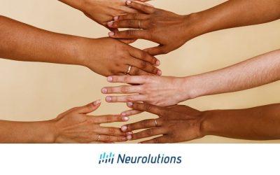
Social interactions may help stroke survivors improve focus and lower both depression and anxiety. Engaging in a creative arts activity may also boost overall neuroplasticity (21). Recent studies have found that being involved in art and music increases cortical plasticity. This is especially true for singing, which strongly activates different areas of the brain (22). Surprisingly, some stroke survivors who are no longer able to speak are still able to sing (23).
For stroke survivors, a stimulating environment that fosters social interactions and maintains a calming atmosphere can be beneficial to staying optimistic about recovering lost cognitive and motor skills while enhancing cortical plasticity. For stroke survivors in rehabilitation centers, environmental and recreational aspects can impact the person’s motivation to engage in therapy.
Top Strategies to Boost Cortical Plasticity After a Stroke: Unleashing Your Brain’s Potential
Engaging in activities like classes and games boosts cortical plasticity, helping stroke survivors maintain cognitive health and potentially reduce dementia.
In addition to participating in rehabilitation services after a stroke, incorporating activities that boost brain health is crucial for achieving optimal recovery. Regular physical exercise, mental stimulation, social interaction, a balanced diet, quality sleep, and stress management all contribute to cortical plasticity.
Stroke survivors are at an increased risk of developing dementia, making it essential to participate in activities that promote overall brain health (24). Combining physical rehabilitation with cognitive stimulation can support both motor recovery and cognitive function, leading to a more comprehensive approach to recovery and reducing the risk of further cognitive decline.
Some activities that may enhance brain health abilities and cortical plasticity are:
-
- Social Interaction in group activities: Regularly engaging in conversations and social activities helps maintain cognitive function and emotional well-being, which are crucial for brain health and recovery.
- Taking classes: Engaging in educational activities stimulates cognitive functions, encourages learning, and promotes social interaction.
- Aerobic Exercise: Activities like walking, swimming, or cycling improve cardiovascular health and enhance brain function, supporting the formation of new neural connections.
- Art and Music Therapy: Creating art or playing an instrument engages multiple brain regions, fostering creativity and aiding in motor and cognitive recovery.
- Passion Projects: Engaging in a “passion project”—an activity or hobby that deeply resonates with a stroke survivor—can be a powerful tool for recovery. Whether it’s gardening, painting, playing music, or any other personally meaningful endeavor, these projects not only foster a sense of purpose and joy but also stimulate the brain in unique ways.
Key Takeaways
Cortical plasticity and neuroplasticity refer to the ability of the brain to form new pathways. In some stroke survivors, this can enable stroke-damaged networks to be replaced by the formation of new pathways that open up communication with other parts of the brain. Rehabilitation programs designed to regain lost cognitive and motor skills also promote the brain’s cortical plasticity which, in turn, can increase the likelihood of recovering lost skills and returning to pre-stroke functioning.
Motivating a stroke survivor to partake in exercises that boost their capabilities in performing ADLs may bridge the gap in their stroke recovery. Since concentration and short-term memory deficits often result from a stroke, engaging in prescribed rehab activities such as OT and/or PT exercises can boost both cortical plasticity as well as motor function. While stroke-induced depression can weaken the motivation to participate in rehabilitation activities, working toward regaining as much independent functioning as possible will boost the brain’s cortical plasticity, creating new pathways that will ultimately lead to an improved quality of life.
References:
- Puderbaugh M, and Emmady PD. (2023). Neuroplasticity. In: StatPearls [Internet]. StatPearls Publishing: Treasure Island, FL. Webpage: https://pubmed.ncbi.nlm.nih.gov/32491743/
- Jawabri KH, and Sharma S. Physiology, Cerebral Cortex Functions. In: StatPearls [Internet]. StatPearls Publishing: Treasure Island, FL. Webpage: https://www.ncbi.nlm.nih.gov/books/NBK538496/
- Koziol LF, Budding D, Andreasen N, et al. (2014). Consensus paper: The cerebellum’s role in movement and cognition. Cerebellum Webpage: https://www.ncbi.nlm.nih.gov/pmc/articles/PMC4089997/
- Riès SK, Dronkers NF, and Knight RT. (2016). Choosing words: Left hemisphere, right hemisphere, or both? Perspective on the lateralization of word retrieval. Ann N Y Acad Sci 1369(1): 111-131. Webpage: https://www.ncbi.nlm.nih.gov/pmc/articles/PMC4874870/
- Jawabri KH, and Sharma S. Physiology, Cerebral Cortex Functions. In: StatPearls [Internet]. StatPearls Publishing: Treasure Island, FL. Webpage: https://www.ncbi.nlm.nih.gov/books/NBK538496/
- Alia C, Spalletti C, Lai S, et al. (2017). Neuroplastic Changes Following Brain Ischemia and their Contribution to Stroke Recovery: Novel Approaches in Neurorehabilitation. Frontiers in Cellular Neuroscience 11: 76. Webpage: https://www.ncbi.nlm.nih.gov/pmc/articles/PMC5352696/
- Puderbaugh M, and Emmady PD. (2023). Neuroplasticity. In: StatPearls [Internet]. StatPearls Publishing: Treasure Island, FL. Webpage: https://pubmed.ncbi.nlm.nih.gov/32491743/
- Alia C, Spalletti C, Lai S, et al. (2017). Neuroplastic Changes Following Brain Ischemia and their Contribution to Stroke Recovery: Novel Approaches in Neurorehabilitation. Frontiers in Cellular Neuroscience 11: 76. Webpage: https://www.ncbi.nlm.nih.gov/pmc/articles/PMC5352696/
- Alia C, Spalletti C, Lai S, et al. (2017). Neuroplastic Changes Following Brain Ischemia and their Contribution to Stroke Recovery: Novel Approaches in Neurorehabilitation. Frontiers in Cellular Neuroscience 11: 76. Webpage: https://www.ncbi.nlm.nih.gov/pmc/articles/PMC5352696/
- Lang CE, Wagner JM, Edwards DF, et al. Effects of task-oriented training on motor recovery in stroke patients: a meta-analysis. Stroke. 2010;41(3):564-570. doi:10.1161/STROKEAHA.109.565402.
- Lin K-C, Chen Y-T, Hsu C-J, et al. Effects of mirror therapy on upper-extremity function and motor recovery in stroke patients: a systematic review and meta-analysis. Neurorehabil Neural Repair. 2011;25(8):723-731. doi:10.1177/1545968311415563.
- Humphries, Joseph B., et al. “Motor Network Reorganization Induced in Chronic Stroke Patients with the Use of a Contralesionally-Controlled Brain Computer Interface.” Brain-Computer Interfaces, vol. 9, no. 3, July 2022, pp. 179–92. DOI.org (Crossref), https://doi.org/10.1080/2326263X.2022.2057757.
- Rustamov, Nabi, et al. “Theta-Gamma Coupling as a Cortical Biomarker of Brain-Computer Interface-Mediated Motor Recovery in Chronic Stroke.” Brain Communications, vol. 4, no. 3, 2022, p. fcac136. PubMed, https://doi.org/10.1093/braincomms/fcac136.
- Hesse S, Schulte-Tigges G, Konrad M, et al. Bilateral arm training: a review of evidence for enhancing motor function and cortical plasticity in stroke patients. Neurorehabil Neural Repair. 2008;22(3):285-291. doi:10.1177/1545968307306523.
- Smania N, Corato E, Fiaschi A, et al. Cognitive-motor dual-task training for gait and balance in patients with stroke: a systematic review and meta-analysis. Stroke. 2010;41(1):216-223. doi:10.1161/STROKEAHA.109.572445.
- Cramer LN, Khatri JM, Zeng PL, Volpe EK, Wolf GL. Constraint-Induced Movement Therapy for Upper Extremity After Stroke: A Systematic Review and Meta-Analysis of Randomized Controlled Trials. Stroke. 2017;48(12):3471-3480. doi:10.1161/STROKEAHA.117.017643. https://pubmed.ncbi.nlm.nih.gov/28154062/
- Laver KE, George S, Thomas S, Deutsch JE, Crotty M. Virtual reality for stroke rehabilitation. Cochrane Database Syst Rev. 2017;11. doi:10.1002/14651858.CD008349.pub3.
- Lohse KR, Lang CE, Boyd LA. Is more better? Using metadata to explore dose-response relationships in stroke rehabilitation. Stroke. 2014;45(7):2053-2058. doi:10.1161/STROKEAHA.114.004695.
- Towfighi A, Ovbiagele B, El Husseini N, et al. (2017). Poststroke Depression: A Scientific Statement for Healthcare Professionals From the American Heart Association/American Stroke Association. Stroke 48: e30–e43. Webpage: https://www.ahajournals.org/doi/full/10.1161/STR.0000000000000113
- Chan CS, Slaughter SE, Jones CA, et al. (2015). Greater Independence in Activities of Daily Living is Associated with Higher Health-Related Quality of Life Scores in Nursing Home Residents with Dementia. Healthcare (Basel) 3(3): 503-518. Webpage: https://www.ncbi.nlm.nih.gov/pmc/articles/PMC4939554/
- Innovative Resources. (March 10, 2023). What we can learn from neuroesthetics – the study of how art changes the brain. Webpage: https://innovativeresources.org/what-we-can-learn-from-neuroesthetics-the-study-of-how-art-changes-the-brain/#:~:text=Any%20type%20of%20creative%20expression,traumatic%20brain%20injuries%20or%20stroke.
- Alain C, Moussard A, Singer J, et al. (2019). Music and Visual Art Training Modulate Brain Activity in Older Adults. Frontiers in Neuroscience 13: 182. Webpage: https://www.ncbi.nlm.nih.gov/pmc/articles/PMC6418041/
- American Stroke Association. (May 4, 2022). The healing power of music for stroke survivors. Webpage: https://www.stroke.org/en/news/2022/05/04/the-healing-power-of-music-for-stroke-survivors
- Kuźma E, Lourida I, Moore SF, et al. (2018). Stroke and dementia risk: A systematic review and meta-analysis. Alzheimers & Dementia 14(11): 1416-1426. Webpage: https://www.ncbi.nlm.nih.gov/pmc/articles/PMC6231970/